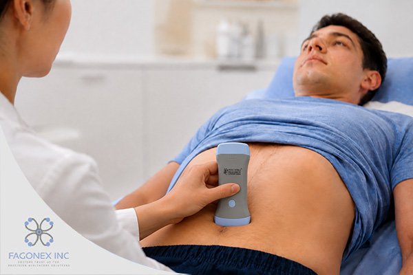
Artificial intelligence is increasingly being incorporated into ultrasound systems, but “AI-powered” can describe very different functions. One device may guide probe movement, another may recognize standard views, and another may automate measurements or flag suspected abnormalities. The key question is whether the specific function is clinically appropriate, validated for its intended users and population, and integrated safely into workflow.
Start With the Intended Clinical Use
First define what the device must do. A system selected for cardiac assessment may require view recognition, ejection-fraction support, and cardiac measurements. A device for vascular access needs high-frequency imaging, responsive needle visualization, and a workflow compatible with sterile procedures. Obstetric, lung, musculoskeletal, and abdominal applications create different requirements.
The FDA’s list of AI-enabled medical devices illustrates that authorization applies to defined devices and intended uses rather than to “AI” as a general capability. The agency states that listed devices have undergone applicable premarket review for safety and effectiveness within their intended use.
A purchasing team should therefore ask what the algorithm is authorized or validated to do—not simply whether the product uses machine learning.
Acquisition Guidance
Obtaining a diagnostic ultrasound image is operator dependent. AI acquisition guidance may provide instructions on probe position, angle, rotation, pressure, or image quality. This can be valuable in training and in focused examinations performed by clinicians who are not expert sonographers.
Research in echocardiography has shown that deep-learning guidance can help novice operators acquire diagnostic transthoracic views. More recent studies report improved view-acquisition success and faster learning with AI-assisted handheld echocardiography, while noting that Doppler-dependent tasks may remain less suitable for frontline automation.
Ask whether guidance works across different body types, disease states, environments, and experience levels. Performance in healthy volunteers may not reflect performance in critically ill patients.
Image-Quality Assessment
Some systems provide a real-time quality score or indicate whether an image is adequate for interpretation. This can reduce the risk that a user stores a technically poor clip and assumes the examination is complete.
A quality score must be linked to the intended task. An image may be adequate for estimating global left ventricular function but inadequate for assessing a subtle valve abnormality. The device should make the scope of its assessment clear.
A review of machine-learning methods for assisted cardiac acquisition found promising performance but noted that many studies lacked sufficient dataset variability. This reinforces the need to examine the populations used for training and validation.
Automated Measurements and Calculations
AI can automate chamber dimensions, bladder volume, obstetric biometry, or estimated ejection fraction. Automation may reduce repetitive work and improve consistency, but clinicians should still be able to inspect the selected borders, landmarks, and source images.
A useful system should show how the result was produced, allow correction, and preserve the original image or cine loop. The organization should know what happens when image quality is inadequate or anatomy falls outside the expected range.
Automated output should support interpretation rather than conceal uncertainty.
Clinical Decision Support
Some devices provide classification or decision-support outputs, which require the highest scrutiny because they may influence diagnosis or treatment.
Before selecting the Best Handheld Ultrasound AI solution for a clinical service, evaluate sensitivity, specificity, external validation, failure modes, and performance across relevant demographic and disease groups. Determine whether the output is intended for screening, triage, measurement, or definitive diagnosis.
The interface should communicate uncertainty, require review of underlying images, and indicate when the algorithm cannot provide a reliable output.
Evidence and External Validation
Vendor demonstrations do not replace clinical evidence. Look for peer-reviewed studies, prospective validation, an appropriate reference standard, and testing outside the developer’s institution.
Important questions include:
- Was the study population similar to your patients?
- Were technically difficult scans included?
- Did performance vary by body habitus or disease severity?
- Was the algorithm tested prospectively?
- Were users representative of the intended operators?
- Were clinically meaningful outcomes measured?
A 2025 systematic review of AI-enhanced breast ultrasound found potential value but also highlighted evidence limitations and the need for stronger validation. Strong performance in one ultrasound task does not establish reliability in another.
Workflow and Interoperability
An AI feature has limited value if it adds delays or creates a separate documentation system. The device should fit into patient identification, image labeling, reporting, storage, and review workflows.
DICOM supports interoperability among imaging devices, PACS, workstations, and other systems. Buyers should verify required export formats, worklists, archive connections, and security controls.
Assess whether processing occurs on-device, on a phone, or in the cloud, because this affects connectivity, latency, data governance, and continuity.
Cybersecurity, Privacy, and Software Updates
Connections to mobile devices, networks, cloud services, or hospital infrastructure create cybersecurity and privacy responsibilities.
FDA guidance addresses managing vulnerabilities during design, deployment, maintenance, and postmarket use. Organizations should understand encryption, authentication, user permissions, update mechanisms, audit logs, data location, and incident-response procedures.
Ask whether software updates can change performance, how changes are validated, and how users are informed.
Hardware Still Matters
AI cannot compensate for unsuitable hardware. Evaluate image quality, probe frequencies, Doppler modes, frame rate, battery life, ergonomics, display compatibility, durability, and cleaning requirements.
A comparison of six handheld devices found that experts prioritized image quality, ease of use, portability, probe size, and battery life. The best AI interface will not solve a mismatch between the transducer and the clinical application.
Choose a Clinical Tool, Not an AI Label
An AI-powered ultrasound device should be selected as a complete clinical system. Its algorithm, hardware, training program, regulatory status, evidence base, interoperability, security, and support must work together.
The most valuable AI functions address a defined limitation without hiding uncertainty or weakening clinical oversight.
A responsible purchase decision begins with the clinical question, examines the evidence behind the specific function, and confirms that clinicians remain able to review the images and make the final judgment.
:: بازدید از این مطلب : 0
|
امتیاز مطلب : 0
|
تعداد امتیازدهندگان : 0
|
مجموع امتیاز : 0


 عضو شوید
 عضویت سریع
 آمار مطالب
 آمار بازدید


